[ad_1]
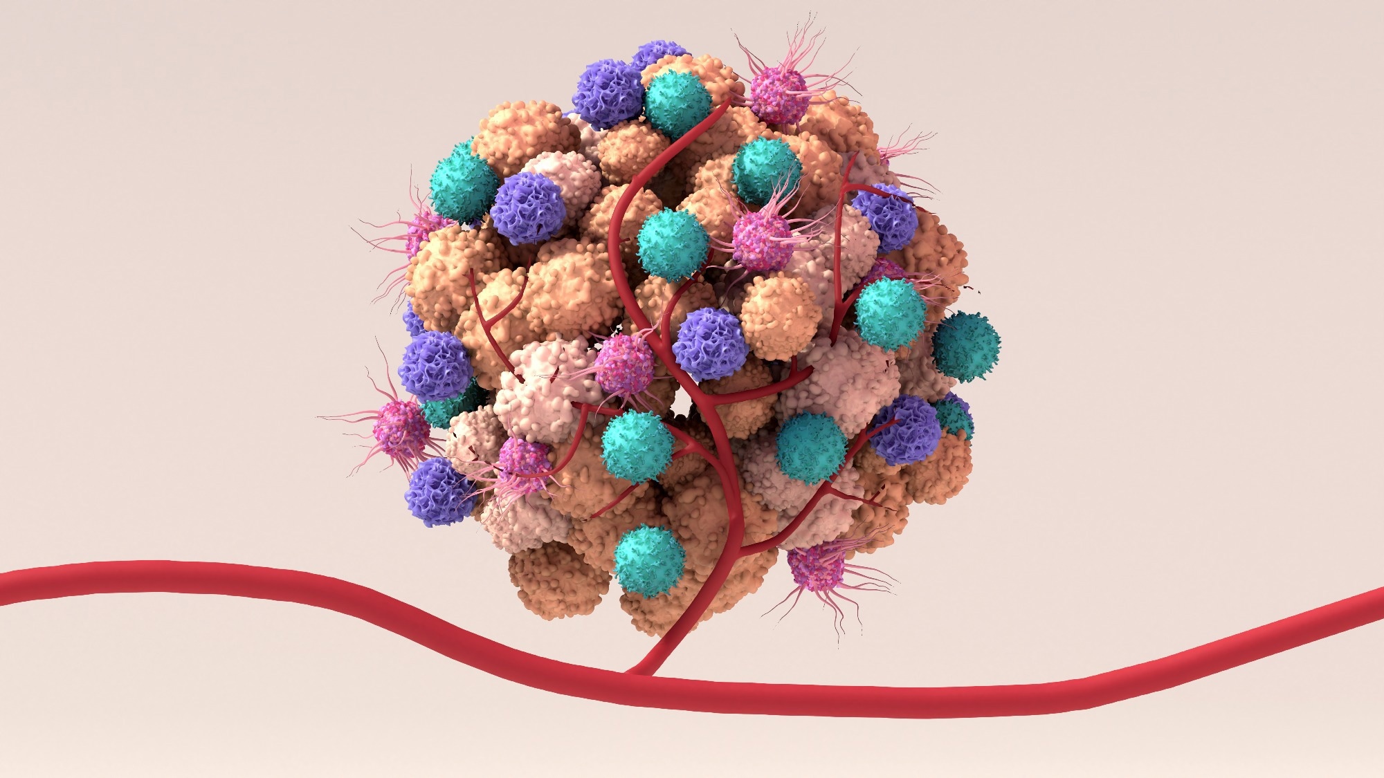
In a current research printed in Cell, researchers investigated whether or not the noticed tumor mobile heterogeneity and structure end result from stochastic and chaotic occasions or whether or not there’s extra coordination within the histopathological chaos of most cancers. They additionally explored mechanisms shaping the extremely difficult tumor panorama.

Both embryology research and oncology analysis actions are aimed toward elucidating tissue development mechanisms; nonetheless, they share restricted conceptual and technical overlap. Morpho-genomic processes and tissue-level signaling are prime mechanisms within the discipline of embryology. Researchers have developed numerous approaches to discover the related mechanisms; nonetheless, cell, genetic, and organic means have been predominantly centered upon, leaving the spatial features of tumor cells under-studied within the discipline of oncology.
About the research
In the current research, researchers analyzed CRCs (colorectal cancers) in three dimensions to evaluate tumor cell heterogeneity and the organizational patterns in tumor cells.
Multiplexed tissue imaging and spatial transcriptomics analyses have been carried out and three-dimensional reconstructions have been analyzed. CRC structure was decided primarily based on constantly occurring transitions starting from the middle of the tumor middle towards the invasive tumor margins. The morphological gradients in mobile conduct and genomic expression have been analyzed.
The tumors confirmed structured structure with coincident gradients in tissue morphology, cell kind distribution, cell perform, and gene expression. Multiple histological phenotypes have been noticed in a tumor. Colorectal cancers have been discovered to resemble morphogenetic patterning concerned within the growth of organs throughout embryogenesis.
Cellular constructions showing as distinct options in two-dimensional photographs have been discovered to be elements of larger three-dimensional options. E.g., perceived tumor budding, showing as tiny and remoted clusters of cells within the two-dimensional photographs, was a large-sized fibrillar protrusion that was detectable solely within the three-dimensional photographs.
The fibrils displayed progressively occurring modifications in mobile proliferation and the expression of signature genes, reminiscent of throughout mobile epithelial-mesenchymal transition (EMT), underpinning that the epithelial-mesenchymal transition happens progressively and never as a binary swap of destiny. Gradually modulated and coherent structure was noticed even within the tumor microenvironment, along with the epithelium of tumors. Tertiary lymph-related constructions appeared as discrete clusters of immunological cells within the two-dimensional photographs. Still, they exhibited interconnected networks with progressively occurring alterations within the composition of the immune cells when examined in three dimensions.
The gradual gradient of functionally distinctive tumor and immunological cells culminated in a number of pathways of T-lymphocyte depend discount amongst morphologically numerous areas of the tumor. The spatial evaluation findings indicated that PD-1 (programmed cell loss of life protein 1)-expressing T lymphocytes, occupying many of the tumor quantity, may be inhibited by PDL1 (programmed loss of life ligand 1)-expressing myeloid cells. Of curiosity, tumor cells located on the budding website expressed PDL1 and will suppress T lymphocyte infiltration on the website.
Conclusions
To conclude, primarily based on the research findings, chaotic stable tumors are, actually, spatially organized tissues from a molecular to tissue scale, indicating that tumors evolve with autonomous patterns of their organic programs. The research findings present a technological development and precious organic insights into the morphogenetic mechanisms and tissue-level signaling pathways amongst tumor cells.
The findings indicated that the spatial orientation of intact tumors is imprinted within the morphological and mobile transitions, various in magnitude and that large-scale spatial assessments have to be carried out to ass the organic variations in tumor samples. Future oncological approaches should combine immunological cell phenotyping and transcriptomic analyses to characterize tumor cell biology additional.
The utilization of histopathological advances to research intact tumor volumes and mixing spatial assays reminiscent of metabolomic and genomic profiling with high-volume data processing and histopathological digitization to enhance the understanding of tumors as difficult but spatially structured lots with organized developmental pathways. It could be easy to research the grade and subtype of tumors; nonetheless, the uncommon areas of the tumor could possibly be of scientific relevance and assist in figuring out tumor recurrence.
TMA (tumor microarrays have to be utilized with warning in drawing organic inferences since TMA areas can’t seize giant three-dimensional options and should not actually consultant of the whole tumor. Orthogonal tissue elimination approaches might overcome the constraints of conventional slide evaluation of tissues and allow the three-dimensional appreciation of tumors as spatially structured lots.
