[ad_1]

Sabine Steidl, RGZM/Burkhard Schillinger, MLZ
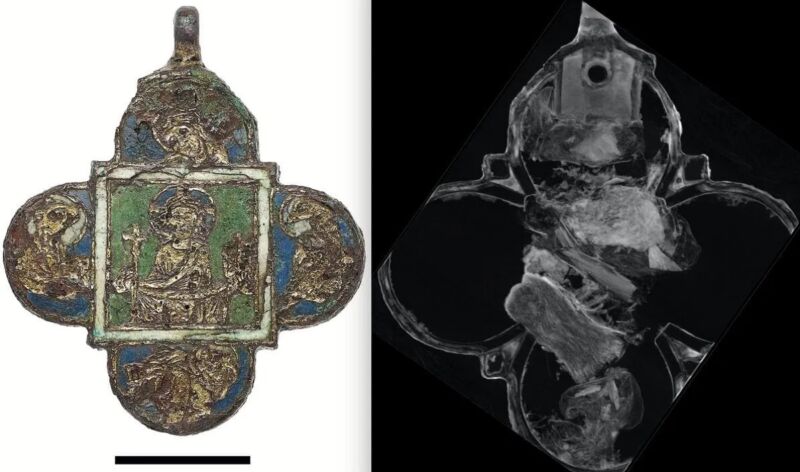
In 2008, archaeologists excavating a medieval refuse pit in Mainz, Germany, found a closely corroded pendant probably made within the late twelfth century. But they have been loath to open the pendant to seek out out what is likely to be inside, lest they injury an already fragile artifact. Now expertise has come to the rescue. Researchers from the Technical University of Munich scanned the pendant utilizing neutron tomography, amongst different strategies, and found it contained bone splinters—probably spiritual relics, i.e., the purported bones of saints. The findings have been printed within the interim assembly of the International Council of Museums-Committee for Conservation (ICOM-CC) Metals Working Group.
Neutron tomography, works a lot the identical manner as X-ray and gamma ray imaging strategies, besides it makes use of a neutron beam. One shoots a beam of radiation on the goal object, and a few elements work together with the pattern whereas others go by way of. The latter collides with an imaging goal to create what’s often called an attenuation sample—primarily a picture of the inside of the pattern. Neutron tomography just isn’t as delicate to the density of supplies as X-ray and gamma ray imaging, and in contrast to these strategies, neutrons work together strongly with very mild components like hydrogen. So some issues simply seen with neutron imaging could also be difficult or unimaginable to see with X-ray imaging (and vice versa).
The methods may be complementary and are particularly helpful for imaging archaeological or paleontological artifacts as a result of they do not injury or destroy the unique object. For occasion, in December 2021, researchers mixed X-ray microtomography—which entails utilizing X-rays to make cross-sections of a bodily object—and neutron tomography to create a extremely detailed 3D mannequin of a 365-million-year-old ammonite fossil from the Jurassic interval, revealing inner muscle groups which have by no means been beforehand noticed. Among different findings, they noticed paired muscle groups extending from the ammonite’s physique, which they surmise the animal probably used to retract itself additional into its shell to keep away from predators.

Sabine Steidl, RGZM
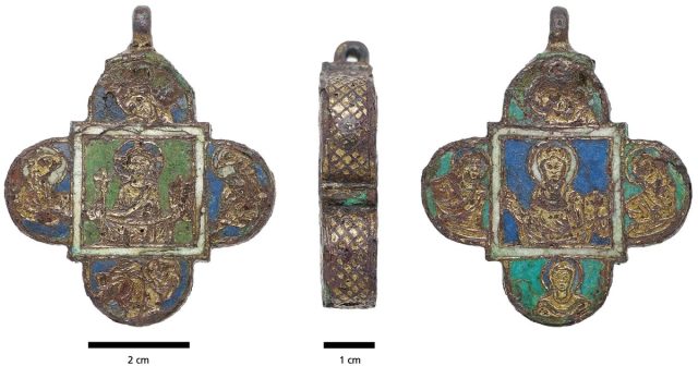
The gold-plated copper pendant in Mainz measures simply 2.4 inches (6 centimeters) excessive and broad, and is within the form of a quatrefoil (a form frequent in conventional Christian symbolism). The back and front are enameled utilizing a method often called champlevé, which entails carving or etching troughs into the floor of a metallic object after which filling them with porcelain enamel. The uncovered parts are gilded, a standard apply in medieval occasions. One facet depicts Jesus, with 4 evangelists pictured within the 4 rounded ends. The different facet options Mary surrounded by 4 feminine saints.
The workforce first analyzed the floor utilizing a mixture of micro-X-ray fluorescence and Raman spectroscopy to determine all the weather current. And infrared spectroscopy revealed a small pattern of beeswax. However, “We couldn’t simply open the trailer and look inside,” mentioned Matthias Heinzel, a restorer at Leibniz-Zentrum für Archäologie (LEIZA), a part of the Technical University of Munich. “The object and, above all, the locking mechanism have been severely damaged by centuries of corrosion, and opening it would mean destroying it irrevocably.”
Using neutron imaging preserved the pendant whereas revealing 5 small reliquary packages of silk and linen holding bone splinters. Heinzel et al. recognized particular person components of the pattern by triggering them with a gamma ray method known as immediate gamma activation evaluation (PGAA). “We can’t say whether or not these bone splinters are from a saint and, if so, which one,” mentioned Heinzel. “Usually relic packages include a strip of parchment indicating the identify of the saint. In this case, nonetheless, we sadly can’t see one.”
The now-fully restored pendant is currently on show on the Mainz State Museum.
